
As a poultry farmer or breeder, understanding the intricacies of fertile egg development is crucial for successful breeding practices. Did you know that from ovulation to hatching, a fertile egg undergoes a series of remarkable transformations? In this comprehensive guide, we’ll take you through each stage of fertile egg development, shedding light on the essential processes that ensure healthy chicks.
We’ll delve into the world of poultry reproduction, explaining how ovulation and fertilization occur, and what happens as the embryo develops inside the egg. Whether you’re a seasoned farmer or just starting out, our expert tips will help you optimize your breeding program and increase hatch rates. By the end of this article, you’ll have gained a deeper understanding of fertile egg development stages, empowering you to make informed decisions about your poultry operation.
Understanding Fertile Eggs
To get a deeper understanding of fertile eggs, let’s dive into the development stages that turn them from ordinary to extraordinary. This process involves several key milestones.
What are Fertile Eggs?
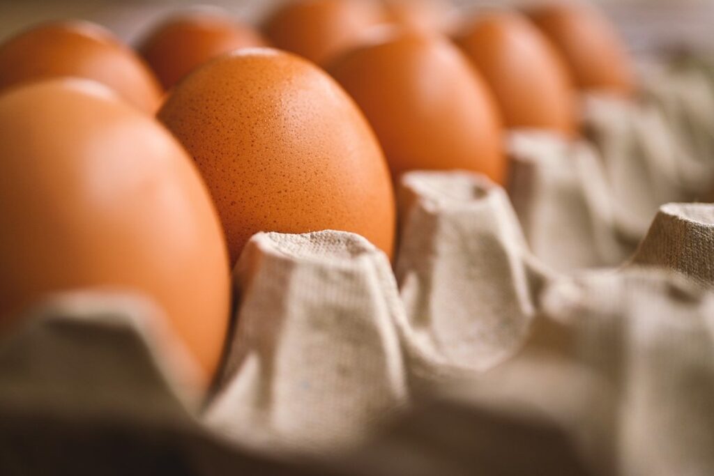
When it comes to poultry breeding, fertile eggs play a crucial role in determining the success of your flock’s reproduction. But what exactly are fertile eggs? In simple terms, fertile eggs are those that have been fertilized by a rooster’s sperm, allowing them to develop into a chick. This process typically occurs when a hen lays an egg and then mates with a rooster, resulting in the release of sperm into the cloaca, where it can fertilize the yolk.
On the other hand, infertile eggs are those that have not been fertilized by a rooster’s sperm. While they may still hatch into chicks through artificial insemination or other methods, natural reproduction is not possible without fertilization. The difference between fertile and infertile eggs lies in their ability to develop into healthy, viable chicks.
As a poultry breeder, it’s essential to recognize the signs of fertile eggs, such as a more robust yolk and a clear or slightly cloudy albumen. By identifying fertile eggs, you can make informed decisions about which birds to breed together, ultimately improving your flock’s overall health and productivity.
Importance of Fertile Eggs in Poultry Breeding
Fertile eggs are a crucial component of poultry breeding, playing a vital role in hatchery production and genetic improvement programs. When it comes to raising healthy and productive birds, the quality of fertile eggs is paramount. For hatcheries, fertile eggs provide a reliable means of producing chicks for commercial farms, research institutions, or backyard flocks.
In genetic improvement programs, fertile eggs enable breeders to introduce desirable traits such as disease resistance, improved growth rates, or enhanced egg-laying capabilities into their flocks. By selecting and breeding from the best lines, farmers can increase the overall performance and efficiency of their poultry operations.
To maximize the benefits of fertile eggs, breeders should focus on producing high-quality hatching eggs with optimal fertility rates. This involves maintaining a healthy flock, monitoring reproductive health, and implementing strict biosecurity protocols to prevent disease outbreaks. By doing so, farmers can ensure that their chicks are raised in an environment conducive to growth and development, ultimately leading to improved productivity and profitability.
Stage 1: Ovarian Follicular Development
Let’s dive into the first stage of fertile egg development, where tiny ovarian follicles begin to form and mature in your ovaries. This is a crucial period for their growth and future success.
The Ovarian Follicle Formation Process
The process of ovarian follicle formation is a complex and intricate one. It begins with the recruitment of oocytes from the primordial follicles, which are formed during fetal development. The primordial follicles contain a dormant oocyte surrounded by flat granulosa cells. As the female grows and matures, certain follicles begin to grow and develop, while others remain dormant.
The selection process for follicle growth is influenced by various factors, including hormonal fluctuations and genetic predisposition. Typically, only around 20-30% of primordial follicles will eventually mature into follicles capable of ovulation. This selection process ensures that only the healthiest and most viable oocytes are chosen to undergo further development.
Once a follicle begins to grow, it enters a phase of rapid proliferation, during which the granulosa cells surrounding the oocyte multiply and differentiate. The growing follicle also becomes responsive to hormones, particularly FSH (follicle-stimulating hormone), which stimulates further growth and maturation.
Growth and Maturation of F Follicles
As F follicles grow and mature in response to hormonal stimuli, they undergo significant changes that ultimately lead to ovulation. This process is triggered by the release of follicle-stimulating hormone (FSH) from the pituitary gland, which stimulates the growth and development of multiple follicles within the ovaries. The dominant F follicle, also known as the leading follicle, receives more intense hormonal stimulation and grows at a faster rate than its counterparts.
As the dominant F follicle continues to mature, it begins to produce estrogen in response to the increasing levels of FSH. This surge in estrogen production causes the follicle to grow even larger and further differentiates into a pre-ovulatory follicle. The increased estrogen also stimulates the growth of the endometrium within the uterus, preparing it for potential implantation of a fertilized egg.
By around day 10-14 of the menstrual cycle, the dominant F follicle has reached its peak size and is now ready to release an oocyte (egg) in response to the luteinizing hormone surge. This marks the onset of ovulation, where the mature egg is released from the follicle and into the fallopian tube, where it can be fertilized by sperm.
Stage 2: Ovum Release and Fertilization
Now that we’ve covered ovulation, let’s dive into what happens next – the release of an egg from the follicle and its potential fertilization by sperm. This is a critical stage in fertilized egg development.
The Process of Ovulation
Ovulation is a complex process where the mature oocyte, also known as an egg cell, is released from the ovarian follicle. This usually occurs around day 14 of a 28-day menstrual cycle, but can vary depending on individual factors such as age and fertility.
In preparation for ovulation, the pituitary gland releases a surge of luteinizing hormone (LH), which stimulates the dominant follicle in one of the ovaries to release its mature oocyte. This process is often accompanied by physical symptoms such as mild pelvic pain or discomfort, breast tenderness, and increased cervical mucus production.
The released oocyte then enters the fallopian tube, where it can be fertilized by sperm within 12-24 hours. If not fertilized, the egg will continue to travel down the fallopian tube and into the uterus, eventually being absorbed or shed during the next menstrual cycle.
It’s essential for women trying to conceive to understand ovulation and its timing, as this increases their chances of getting pregnant. Women can track their ovulation using basal body temperature (BBT) charting, fertility apps, or by monitoring their cervical mucus changes.
Fertilization Mechanisms
When a mature ovum is released from the follicle and enters the fallopian tube, it’s swept by tiny hair-like projections called cilia towards the uterine end. Here, it awaits potential fertilization. Fertilization occurs when a sperm penetrates the outer layer of the ovum, breaking through its zona pellucida.
The process begins with sperm capacitation – a transformation that allows them to bind to and penetrate the egg’s outer layer. This is facilitated by chemical signals from the female reproductive tract. The first sperm to successfully penetrate the zona pellucida fuses with the ovum’s plasma membrane, triggering a series of calcium ion influxes.
The resulting zygote undergoes rapid cell division and development. As this occurs, it begins to absorb nutrients from surrounding fluid, gradually increasing in size and complexity. A critical factor in successful fertilization is timing – sperm must penetrate within 12-24 hours after ovulation for optimal chances of successful conception.
Cleavage Stages: Early Embryogenesis
As we’ve discussed ovum release and fertilization, let’s dive into the fascinating world of early embryonic development. After successful fertilization, the zygote begins to divide through a process called cleavage. This rapid cell division occurs over several hours, resulting in a group of cells that will eventually form the embryo.
Cleavage stages can be broadly categorized into several phases:
- Mosaicism: The first few divisions are uneven, resulting in different-sized cells with varying amounts of cytoplasm.
- Holoblastic cleavage: The entire egg is divided into two equal parts with each new cell having an equal amount of cytoplasm.
- Meroblastic cleavage: Only a portion of the egg undergoes division.
As these divisions continue, the embryo begins to compact and form a blastocyst – a hollow ball of cells that will eventually give rise to the fetus. This critical stage, usually around 5-6 days post-fertilization, marks the transition from an early embryo to a more complex organism.
Stage 3: Pre-Implantation Development
As we move forward in our journey through fertile egg development, let’s dive into the crucial pre-implantation stage, where key processes prepare for implantation. This is a critical period of rapid growth and transformation.
Morula Formation and Blastulation
As we’ve discussed the formation of the zygote and its transition into the compacted mass of cells known as the morula, it’s now time to explore the next key stage: blastulation. During this period, significant changes occur that lay the groundwork for implantation.
The morula starts to undergo a process called cavitation, where fluid-filled spaces within the cell cluster begin to form. This marks the beginning of the transition into the blastocyst stage. As the cells continue to compact and differentiate, the morula’s spherical shape gives way to a more elongated form, with one pole becoming distinct from the other.
The formation of the trophoblast layer is another crucial development during this period. This outer layer will eventually give rise to the placenta and surrounding tissues that support fetal growth. Meanwhile, the inner cell mass (ICM) begins to take shape, comprising cells that will go on to form the embryo itself.
As you can see, the transformation from morula to blastocyst is a complex yet crucial process in fertile egg development. Understanding these early stages can help us better appreciate the intricate mechanisms at play as an embryo develops into a healthy fetus.
Implantation Preparation
As we reach the final stages of pre-implantation development, significant changes occur within the embryonic structure to prepare it for implantation into the uterus lining. Around 6-7 days post-fertilization, the embryo undergoes a process called compaction, where its cells become tightly packed and begin to take on a more compact form.
This transformation is crucial as it enables the embryo to withstand the mechanical stresses of implantation. The embryoblast layer begins to differentiate into two distinct groups: the inner cell mass (ICM) and the trophoblast layer. The ICM will eventually give rise to the fetus, while the trophoblast layer develops into the placenta.
In preparation for implantation, the embryo’s surface also undergoes a transformation, becoming more receptive to attachment with the uterus lining. This process is facilitated by the production of adhesion molecules on the embryonic surface, which enable it to bind securely to the uterine epithelium. These adaptations ensure that when the embryo finally implants into the uterine lining, it can establish a stable connection and initiate nutrient and gas exchange.
In essence, the changes occurring during this stage are critical for successful implantation and subsequent fetal development. By understanding these processes, you’ll appreciate the intricate complexities of embryonic growth and development.
Stage 4: Gastrulation and Organogenesis
Now that we’ve covered the early stages of egg development, let’s dive into the fascinating process of gastrulation and organogenesis, where your chick’s body starts to take shape. This is a crucial time for growth and differentiation.
The Beginnings of Gastrulation
As we reach the gastrulation stage, you’ll notice a significant transformation taking place within the fertilized egg. This critical period marks the transition from a blastula to a gastrula stage, where the embryo’s basic body structure begins to take shape.
Gastrulation starts around 16-24 hours after fertilization, and it’s a complex process that involves cell migration and differentiation. At this point, the blastula’s single layer of cells starts to thicken and fold inwards, forming three primary germ layers: ectoderm, mesoderm, and endoderm.
These germ layers will eventually give rise to all the tissues and organs found in the body. For instance, the ectoderm differentiates into the central nervous system and skin, while the mesoderm develops into muscles, bones, and blood vessels.
As gastrulation progresses, you’ll notice the formation of the archenteron, a precursor to the digestive system. This is an essential step in the development process, as it sets the stage for organogenesis – the creation of more complex organs and body systems. With a solid understanding of gastrulation, you can appreciate the intricate processes at play during early embryonic development.
Organ Formation and Morphogenesis
During organogenesis, embryonic tissues undergo a complex series of transformations to give rise to various organs and systems. This process is essential for the development of a functional and viable organism. As gastrulation comes to an end, the embryo begins to organize itself into distinct layers, each containing specific groups of cells with different fates.
The ectoderm layer, for instance, gives rise to the nervous system, skin, and sensory organs, while the mesoderm develops into muscles, bones, and connective tissue. The endoderm, meanwhile, forms the lining of internal organs such as the gut, liver, and lungs. Through a series of intricate cellular interactions and migrations, these germ layers eventually give rise to fully formed organs.
For example, in chicken embryos, the development of the gut is initiated by the invagination of the endodermal cells into the mesoderm layer, followed by the differentiation of these cells into specific types of intestinal epithelial cells. Similarly, the formation of the heart involves the convergence of cardiac progenitor cells from the splanchnic mesoderm to form a single tube that eventually divides into separate chambers.
Understanding the mechanisms behind organogenesis can provide valuable insights for researchers working on regenerative medicine and tissue engineering. By studying how embryonic tissues give rise to functional organs, scientists can develop novel approaches for repairing damaged tissues and organs in adults.
Stage 5: Embryogenesis and Hatching
Now that your egg is developing its embryo, let’s dive into the fascinating process of embryogenesis, where life begins to take shape. This stage sets the stage for hatching.
Embryo Development in the Uterus
As we continue through the embryogenesis and hatching stage, let’s dive into the fascinating process of embryo development within the uterus. After implantation, the fertilized egg undergoes a series of rapid transformations, eventually forming a complex structure known as a blastocyst.
During this period, which spans around 6-7 days post-fertilization, the embryo experiences significant morphological changes. The inner cell mass (ICM) begins to differentiate into two distinct groups: the epiblast and the hypoblast. The epiblast will eventually give rise to all three germ layers – ectoderm, mesoderm, and endoderm – while the hypoblast contributes to the formation of extra-embryonic tissues.
As the embryo grows, it begins to fold in upon itself, forming a structure known as a bilaminar disc. This folding process is crucial for establishing the embryo’s anterior-posterior axis, setting the stage for future development and organogenesis. By around 7-8 days post-fertilization, the embryo has reached the blastocyst stage, marking a critical milestone in its journey towards hatching and implantation into the uterine wall.
Preparation for Hatch
As you approach the final stages of embryogenesis, the developing chick is preparing for its grand entrance into the world. Lung formation is a critical process that enables the chick to breathe outside the egg. At around 17-18 days of incubation, lung development begins with the differentiation of the respiratory epithelium and the formation of the air sacs.
The chick’s lungs are initially non-functional and gas exchange occurs through the allantois, a yolk sac membrane that provides oxygen and removes waste products. However, as the chick grows, the allantois starts to degenerate, and the lungs take over respiratory functions. By 19-20 days, lung development is complete, and the chick’s first breaths are imminent.
As you prepare for hatching, make sure the incubator temperature is at a safe range (99°F – 100°F) and maintain humidity levels between 50-60%. Ensure proper ventilation to prevent carbon dioxide buildup. Monitor the egg closely, and be prepared for pipping, which usually occurs within 24 hours of the final lung formation stage.
Post-Hatch Care and Handling
Now that you’ve successfully hatched your fertile eggs, it’s time to learn how to care for these delicate chicks and ensure they thrive in their new environment.
Initial Care and Nutrition
After your fertile eggs hatch, it’s essential to provide proper care and nutrition for the newly emerged chicks. This critical period is known as brooding, where you’ll need to ensure a warm and safe environment for their growth and development.
Maintain a consistent temperature between 90-100°F (32-38°C) during the first week of life. You can achieve this using heat lamps or heat mats placed under a shallow dish filled with water. This will help regulate their body temperature, as they rely on external sources to keep warm.
A starter feed or chick starter mash should be introduced within 24 hours after hatching. Provide free-choice access to fresh water at all times and make sure the feeding trough is kept clean. Feed 20% protein starter feed for the first four weeks of life, gradually increasing the amount as they grow. Monitor their intake and adjust feeding schedules accordingly.
Keep a close eye on your chicks’ droppings, ensuring they’re well-formed and brown in color. This will indicate proper digestive health and functioning. Monitor the temperature, humidity levels, and light exposure to prevent any potential health issues. With proper care and attention, your newly hatched chicks will develop into healthy and robust birds.
Housing and Management Considerations
When it comes to post-hatch care and handling, housing and management considerations are crucial for the health and well-being of newly hatched chicks. To minimize stress and prevent disease, it’s essential to provide a safe and clean environment.
First and foremost, ensure that the brooder or chick enclosure is properly ventilated to prevent ammonia buildup from droppings. A good rule of thumb is to maintain a temperature range of 90-95°F (32-35°C) for the first week, with gradual reductions over time. Keep in mind that chicks need access to fresh air and adequate space to move around.
Implementing biosecurity measures is also vital. Disinfect all equipment and surfaces regularly, and avoid overcrowding to prevent the spread of disease. Consider using a quarantine area for new arrivals or chicks showing signs of illness. By prioritizing housing and management considerations, you’ll be well on your way to raising healthy, happy chicks.
Frequently Asked Questions
How do I ensure the optimal temperature for fertile egg incubation?
Maintaining a consistent temperature is crucial for successful hatching. Aim for an average temperature of around 99-100°F (37°C) during the first 24 hours after laying, and then gradually decrease it to about 98°F (36°C) by day 10. This precise control will promote healthy chick development.
Can I use artificial incubation methods if I have a small flock?
Yes, artificial incubators are an excellent option for small-scale poultry breeding. They provide a controlled environment, allowing you to monitor and adjust temperature, humidity, and turning cycles with precision. This is particularly useful when working with limited space or numbers.
How can I identify potential fertility issues in my roosters?
Keep an eye on your rooster’s behavior, ensuring he has ample access to hens for mating. Also, regularly inspect his sperm quality through a process called “semen collection and analysis.” This will help you pinpoint any fertility problems early on and make necessary adjustments.
What are the most common causes of low hatch rates?
Low hatch rates can be attributed to various factors such as poor egg handling, incorrect temperature or humidity levels during incubation, inadequate turning, or even underlying health issues within the flock. Regularly monitoring these variables will help you pinpoint potential problems before they become major concerns.
Can I reuse fertile eggs in subsequent breeding cycles?
No, it’s not recommended to reuse fertile eggs for future breeding purposes. Fertile eggs are designed for a single cycle of incubation and hatching; attempting to reuse them may result in reduced fertility or even complete sterility due to the physical stress on the embryo.