
As you crack open an egg to prepare for cooking, have you ever wondered about the tiny miracle unfolding inside? Chicken embryonic development is a complex process that’s crucial for understanding how our feathered friends come into being. From the initial division of cells to the eventual hatching of a fluffy chick, this journey is marked by critical milestones and factors that affect success. Genetic influences play a significant role in determining traits such as eggshell color and fertility, making chicken embryonic development a fascinating area of study. In this article, we’ll delve into the intricate process of cleavage, gastrulation, and organogenesis, exploring the key events and factors that shape the development of a chicken embryo from fertilization to hatching.
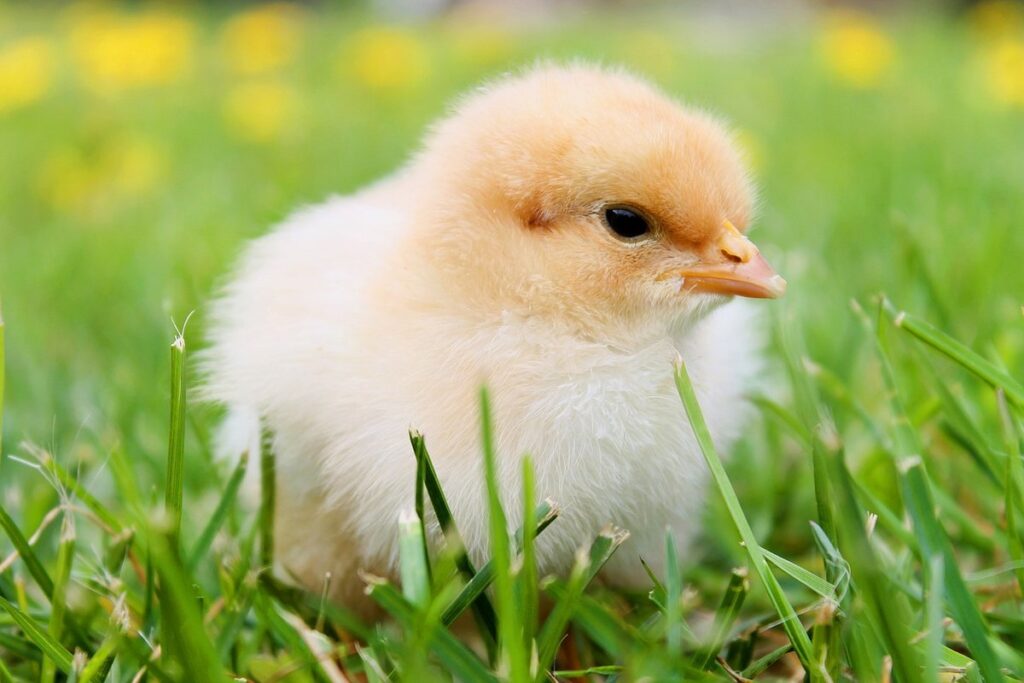
Stage of Pipping and Hatching
As we continue our journey through chicken embryonic development, let’s dive into the exciting stages where beaks start to crack and tiny wings begin to stretch. We’re almost at hatch!
The Eggshell’s Role in Embryo Protection
The eggshell plays a crucial role in protecting the embryo from external factors during incubation. It’s like a fortress that shields the developing chick from harm. One of its primary functions is to regulate temperature and humidity levels, keeping them within a narrow range that’s perfect for growth.
Imagine you’re placing an egg in a warm oven or under direct sunlight – it wouldn’t take long for the embryo to overheat and suffer damage. That’s why the eggshell helps maintain a stable temperature between 99°F and 100°F (37°C to 38°C). It also prevents moisture loss, keeping the embryo hydrated throughout incubation.
The eggshell’s structure is designed to withstand external pressures while still allowing gases like oxygen and carbon dioxide to pass through. This ensures that the embryo gets the necessary nutrients for growth without being exposed to harsh conditions outside.
In fact, studies have shown that eggs with strong, intact shells tend to hatch at a higher rate than those with weakened or cracked shells. So, if you’re incubating eggs at home, make sure to handle them gently and keep them away from drafts to ensure the eggshell remains intact and functional throughout the incubation period.
Critical Factors Affecting Pipping Success
Temperature control is critical during pipping and hatching. A temperature range of 99-100°F (37.2-37.8°C) is ideal for this stage. However, it’s essential to maintain a consistent temperature, as fluctuations can cause stress to the embryo.
Humidity also plays a crucial role in successful pipping and hatching. Maintain a humidity level between 50-60% to prevent excessive moisture loss. Monitor your incubator’s hygrometer closely to ensure accurate readings.
Air circulation is equally vital for healthy development during this stage. Ensure good air exchange by following the manufacturer’s instructions for air vents or adjusting the ventilation settings on your incubator. A well-ventilated environment promotes gas exchange and prevents the buildup of CO2, which can lead to respiratory distress.
Adequate temperature, humidity, and air circulation will significantly increase the chances of successful pipping and hatching. Be cautious not to over-ventilate, as this can cause excessive moisture loss and negatively impact development. Regularly check your incubator’s settings and monitor your eggs for signs of stress or discomfort.
Developmental Milestones Before Hatching
As you await the arrival of your chicks, it’s essential to understand what’s happening inside the egg during this critical period. Developmental milestones before hatching are a complex series of events that ultimately lead to the emergence of your new chicks.
About 18-20 days into incubation, the embryo undergoes a significant transformation – the formation of the beak and legs. This is a crucial stage as it sets the foundation for the chick’s ability to pip (break through the shell). You’ll notice the beak starting to develop, followed by the appearance of tiny claws.
As development progresses, the embryo’s growth accelerates, and you may start noticing movement within the egg. This can be an exciting time for breeders, but it’s essential to remain patient and let nature take its course. Around 21-22 days into incubation, the embryo’s eyes begin to form, and you may even see a faint light reflecting off the developing retina.
Remember that every chick develops at its own pace, so it’s crucial to monitor temperature and humidity levels closely during this period. A consistent environment will help ensure your chicks emerge strong and healthy.
Stages of Embryogenesis in Chickens
Let’s take a closer look at how chickens develop from tiny embryos to adorable chicks, and break down each crucial stage in their embryonic journey. From fertilization to hatching, it’s an incredible process.
Cleavage Stage (0-24 hours)
At the start of embryonic development in chickens, we have the cleavage stage, which spans from 0 to 24 hours after fertilization. During this period, the process of zygote formation and initial cell divisions take place. Let’s dive into the details.
After fertilization, the sperm penetrates the egg yolk, leading to the fusion of male and female gametes, resulting in the formation of a zygote. The zygote then undergoes its first cell division, known as cleavage, within the germinal disc. Cleavage is characterized by the rapid division of cells without significant growth.
The initial cell divisions occur at a rate of approximately 15 minutes per cell cycle. This results in an increase in the number of blastodermal cells from a single zygote to around 16-32 cells within 24 hours. The embryo’s cells become polarized, with distinct areas forming for the germ layers – ectoderm, endoderm, and mesoderm – which will eventually give rise to various tissues and organs.
The cleavage stage is crucial in establishing the foundation for subsequent developmental processes, including gastrulation, organogenesis, and eventual hatching. Understanding this initial phase of embryonic development helps in managing breeding programs, improving hatch rates, and enhancing overall chicken health.
Morula Formation and Blastulation (24-72 hours)
As we move forward in the embryonic development of chickens, between 24 to 72 hours after fertilization, the morula stage sets the groundwork for further growth. The morula is a cluster of 16-32 cells that have undergone compaction, where they start to stick together and lose their individual cell membranes. This process increases the surface area for nutrient and waste exchange, providing essential energy and resources for the embryo.
Blastulation begins when the morula undergoes cavitation – the formation of a fluid-filled cavity within the cluster of cells. The resulting blastocyst consists of two distinct layers: the inner cell mass (ICM) and the trophectoderm. The ICM gives rise to the fetus, while the trophectoderm differentiates into extra-embryonic membranes such as the amnion, chorion, and allantois.
During this period, it is essential for breeders to maintain optimal environmental conditions – precise temperature control, sufficient moisture, and proper aeration. This helps ensure that embryos develop at an ideal rate and are less susceptible to stress or disease. Proper incubation techniques can significantly influence the success of embryonic development in chickens.
Gastrulation and Organogenesis (3-6 days)
During the 3-6 day period of embryonic development in chickens, two pivotal processes occur: gastrulation and organogenesis. Gastrulation marks a significant transition from a blastula to a gastula stage, where the embryo’s cells begin to organize into three primary germ layers: ectoderm, endoderm, and mesoderm. These layers will eventually give rise to all tissues and organs in the developing chick.
Organogenesis, which follows gastrulation, is the formation of organs from these germ layers. This process involves a complex series of cell movements, differentiations, and interactions that result in the development of specific organ systems, including the heart, lungs, liver, and limbs. During this stage, major tissue differentiation occurs as cells begin to adopt their final forms and positions.
For example, neural crest cells migrate from the ectoderm to form cranial cartilage, facial bones, and parts of the nervous system. Meanwhile, endodermal cells give rise to the lining of the digestive tract, respiratory tract, and other internal organs. Understanding these processes is crucial for poultry breeders and researchers seeking to optimize embryonic development and improve chick viability.
Proliferation and Differentiation of Embryonic Tissues
Let’s dive into the complex process of how different tissues in a chicken embryo begin to proliferate and differentiate during development. We’ll explore the key stages that shape these cells into functioning parts of the body.
Neurulation: The Formation of the Central Nervous System
As we explore the fascinating world of chicken embryonic development, it’s essential to delve into the intricate process of neurulation, which marks the formation of the central nervous system. This critical stage occurs around 18-24 hours after fertilization and sets the foundation for the chick’s cognitive functions.
The neural tube, a flat plate of cells, begins to fold in on itself, eventually forming the brain and spinal cord. As this process unfolds, a series of intricate folds and clefts emerge, ultimately giving rise to distinct regions within the central nervous system. The rostral (front) portion develops into the forebrain, while the caudal (rear) section becomes the hindbrain.
The neural tube’s closure is crucial, as it allows for the proper development of motor neurons, sensory neurons, and glial cells that support these vital components. Any disruption during this sensitive period can lead to developmental abnormalities or even neurological disorders later in life. Therefore, understanding neurulation is essential for grasping the complex mechanisms driving chick embryonic development.
As we continue our journey through the chicken embryo’s development, keep in mind the significance of this early stage in shaping the chick’s nervous system.
Cardiac Development and Blood Vessel Formation
As we delve into the world of chicken embryonic development, it’s essential to understand the intricacies of cardiac development and blood vessel formation. The process begins around day 2-3 of incubation when the cardiac precursor cells start to migrate towards the lateral plate mesoderm. These cells will eventually form the linear heart tube, which then undergoes a series of complex movements known as looping, bending, and twisting.
As the heart tube loops, it differentiates into distinct chambers – the atria and ventricles. The blood vessels, including the arteries and veins, begin to form through a process called vasculogenesis, where endothelial cells sprout from the mesoderm to line up with the cardiac tubes. This is crucial for the delivery of oxygenated blood to the developing embryo.
It’s fascinating to note that, by day 5-6, the heart starts pumping blood through its chambers and vessels, marking a significant milestone in embryonic development. However, this process can be influenced by various factors such as temperature, nutrition, and genetics. Understanding these intricacies is crucial for optimizing hatching rates and healthy chick growth.
In practical terms, breeders and researchers can take steps to support optimal heart development by maintaining precise incubation conditions, monitoring embryo growth, and selecting breeding stock with desirable traits. By doing so, they can increase the chances of healthy chicks and reduce mortality rates.
Muscle and Skeletal System Differentiation
As we delve into the fascinating world of chicken embryonic development, let’s take a closer look at how muscle and skeletal systems differentiate. The process begins with the migration of mesodermal cells to the somites, which will eventually give rise to muscles and bones.
Muscle cell migration is a complex process that involves the coordinated movement of cells to their specific destinations. This is achieved through a series of intricate signaling pathways that regulate cell adhesion, migration, and differentiation. Once at their destination, these precursor cells undergo a series of morphological changes to become myoblasts, which then fuse together to form muscle fibers.
Meanwhile, chondrogenesis is taking place in the adjacent somites. Here, mesenchymal cells differentiate into cartilage-producing cells called chondrocytes. These cells secrete a cartilaginous matrix that provides structural support and eventually gives way to bone ossification. As the embryo grows, this cartilage is gradually replaced by bone tissue through a process called endochondral ossification.
It’s essential to note that the proper alignment of these processes is crucial for the formation of functional muscles and bones. Any disruptions can lead to developmental abnormalities or even congenital conditions in chicks.
Sensory Organ Development in Chickens
Let’s take a closer look at how the sensory organs develop inside the egg, from eyes to ears and everything in between.
Eye and Vision Development
As we explore the development of sensory organs in chickens, let’s take a closer look at eye and vision development. This process is incredibly complex and fascinating, involving the formation of multiple layers and the maturation of specialized cells.
The lens, which focuses light onto the retina, begins to form around day 6-7 of incubation. It starts as a thickened layer of cells and gradually becomes more transparent, allowing for clear vision. The retinal layers also begin to take shape during this period, with neurons and photoreceptors starting to differentiate.
Around day 12-14, the retina undergoes rapid growth, with the formation of multiple cell layers. This is when the rods and cones – the two types of photoreceptor cells responsible for vision – start to mature. Rods are more sensitive to low light levels, while cones are better suited for color vision and high-acuity tasks.
By day 18-20, the retina has reached its final form, with all cell layers fully differentiated. The eye is now capable of detecting light and forming images, although it’s not yet clear or focused. As the embryo develops further, the lens continues to mature, allowing for clearer vision.
Ear and Auditory System Formation
During embryonic development, the formation of the ear and auditory system is a complex process that involves multiple stages. Around 3-4 days after fertilization, the otic placode forms on the side of the embryo’s head, eventually giving rise to the inner ear structures. As the embryo grows, the otocyst differentiates into three main parts: the vestibular (responsible for balance), cochlear (responsible for hearing), and auditory nerve.
By around 5-6 days, the tympanic membrane begins to form, separating the outer ear from the middle ear cavity. This delicate process requires precise regulation of genes involved in cell migration and differentiation. Abnormalities in this process can lead to defects in ear development. The auditory nerve, responsible for transmitting sound signals to the brain, starts to myelinate around 7-8 days after fertilization. Proper myelination is crucial for normal hearing function.
It’s worth noting that disruptions in ear and auditory system development can have significant consequences on a chick’s ability to hear and navigate its environment effectively.
Nutrition and Energy Metabolism in Embryos
As we explore how chicken embryos grow and develop, let’s dive into the crucial role of nutrition and energy metabolism that fuels their rapid growth. This is where tiny cells become fully formed chicks.
Yolk Sac and Amniotic Fluid’s Role in Nutrient Delivery
As the embryo develops inside the egg, it relies heavily on the nutrients stored in the yolk sac for growth and energy. The yolk sac serves as a reservoir of proteins, carbohydrates, fats, vitamins, and minerals that are essential for the embryo’s development. However, these nutrients need to be transferred from the yolk sac to the embryo through the amnion, a membrane that surrounds the embryo.
This transfer occurs through a process called diffusion, where the nutrients in the yolk sac move freely into the surrounding fluid, which is then absorbed by the embryo. The amnion plays a crucial role in this process, as it provides a barrier between the embryo and the yolk sac while allowing for nutrient exchange to occur.
In essence, the amniotic fluid acts as a medium that facilitates the transfer of nutrients from the yolk sac to the embryo. This process is essential for the healthy development of the embryo, ensuring that it receives all the necessary nutrients for growth and energy production.
Liver Development and Lipid Storage
The liver plays a crucial role in chicken embryonic development, particularly when it comes to nutrition and energy metabolism. During this critical period, the liver’s primary function is to process and store nutrients for growth and development.
In terms of glycogen storage, the liver acts as a reservoir, storing excess glucose derived from the mother’s feed or the chick’s own yolk sac in the form of glycogen. This complex carbohydrate is broken down into glucose when needed by the embryo, providing a readily available source of energy. In fact, studies have shown that glycogen reserves can support embryonic development for up to 24 hours after hatching.
Lipid metabolism is also essential during this period, as the liver synthesizes and stores lipids from yolk sac components or maternal feed. This stored lipid reserve is gradually mobilized as the embryo grows, providing a vital source of energy for critical developmental processes. Proper nutrition and energy management are therefore crucial to ensure optimal liver development and function in chicken embryos.
Genetic Factors Influencing Embryonic Development
Let’s take a closer look at the genetic factors that play a crucial role in shaping the early stages of chicken embryonic development, from fertilization to hatching.
Genome Regulation and Transcriptional Activation
When it comes to regulating embryonic development in chickens, genes play a pivotal role. The process begins with genome regulation, where specific genes are activated or suppressed depending on the stage of development. This is achieved through transcriptional activation, which involves the binding of transcription factors to DNA.
Transcription factors are like “on/off switches” for gene expression. They determine which genes should be turned on or off at a particular time during embryonic development. For example, the Hox family of transcription factors regulates limb patterning and morphogenesis in chicken embryos. By controlling the expression of these genes, cells can differentiate into specific tissues and organs.
Key players like SOX2 and OCT4 are crucial for maintaining pluripotency in chicken embryonic stem cells. These transcription factors work together to ensure that stem cells retain their ability to differentiate into various cell types. Understanding how these transcription factors interact with each other and the genome will help us better comprehend the complex mechanisms governing embryonic development. By deciphering these regulatory networks, researchers can develop targeted interventions to improve poultry breeding programs and reduce developmental abnormalities in chicken embryos.
Mutations Affecting Fetal Development
Genetic mutations can significantly impact normal fetal development in chickens. When it comes to embryonic development, the intricate process of gene expression and protein synthesis is crucial for proper growth and differentiation. A mutation affecting a key developmental gene can lead to abnormal morphogenesis, resulting in congenital anomalies or even embryonic lethality.
For example, the Eda gene plays a vital role in feather formation during chick embryogenesis. Studies have shown that mutations in this gene can result in various developmental abnormalities, including extra toes and beak deformities (Yang et al., 2015). Similarly, mutations affecting the Hox genes, responsible for patterning and morphogenesis, can lead to altered limb development and other congenital anomalies.
Understanding the effects of genetic mutations on fetal development is crucial for poultry breeders. By identifying potential genetic issues early in the breeding process, they can make informed decisions about which birds to use as parents, ultimately reducing the likelihood of developmental abnormalities in their offspring.
Some key takeaways from this discussion include:
• Genes such as Eda and Hox are critical for normal embryonic development
• Mutations affecting these genes can lead to congenital anomalies or embryonic lethality
• Understanding genetic mutations is essential for poultry breeders to make informed breeding decisions
While we cannot completely eliminate the risk of genetic mutations, being aware of potential risks and taking proactive steps to identify and mitigate them can go a long way in producing healthy, normal chicks.
Frequently Asked Questions
How Can I Apply the Knowledge of Chicken Embryonic Development to Improve My Hatch Rate?
By understanding the intricacies of chicken embryonic development, you can make informed decisions about breeding programs, nutrition, and incubation conditions to boost your hatch rate. For example, recognizing the importance of proper temperature regulation during incubation can help you avoid overheating or underheating issues that may impact embryo development.
What Are Some Common Mistakes People Make When Caring for Chickens During Incubation?
One common mistake is neglecting to maintain a consistent temperature and humidity level within the incubator. This can lead to delayed hatching, embryonic mortality, or even failure to hatch altogether. Regularly monitoring and adjusting these conditions according to your specific chicken breed’s needs will help you avoid such mistakes.
Can I Use the Information on Chicken Embryonic Development to Improve My Breeding Program?
Yes! Understanding the genetic factors that influence traits like eggshell color and fertility can be invaluable for selecting breeding stock that will produce offspring with desirable characteristics. By incorporating this knowledge into your breeding program, you can make informed decisions about which birds to breed together, potentially leading to healthier, more productive flocks.
How Can I Ensure Proper Nutrition During Embryogenesis?
Proper nutrition is crucial during embryogenesis. Ensuring adequate yolk sac and amniotic fluid levels is essential for delivering necessary nutrients and energy to the developing embryo. Maintaining a clean and well-ventilated incubator can also help prevent bacterial or fungal contamination, which may compromise nutrient delivery.
What Are Some Signs That an Embryo May Be at Risk of Failure to Hatch?
Keep an eye out for delayed pipping (the time it takes for the chick to break through the eggshell), changes in egg weight or appearance, and unusual incubation periods. If you notice any of these signs, consult with a veterinarian or poultry expert to assess the situation and take corrective action if necessary.